Exposure to asbestos can affect risk of malignant mesothelioma.

Most people with malignant mesothelioma have worked or lived in places where they inhaled or swallowed asbestos. After exposure to asbestos, you usually spend much time until a malignant mesothelioma is formed. Living together with a person who works near asbestos also is a risk factor for malignant mesothelioma.
The signs and symptoms of malignant mesothelioma include difficulty breathing and pain under the rib cage.
Sometimes the cancer makes accumulating fluid in the chest or abdomen. The signs and symptoms can be produced from liquid, a malignant mesothelioma or other conditions. Check with your doctor if you experience any of the following symptoms:
Shortness of breath.
Cough.
Pain under the rib cage.
Abdominal pain or swelling of the same.
Nodules in the abdomen.
Constipation.
Problems with blood clots (blood clots that form when they must not do it).
Loss of weight without known reason.
Feeling very tired.
To detect (find) and diagnose malignant mesothelioma, tests are used to examine the inside of the chest and abdomen.
In some cases, it is difficult to differentiate malignant mesothelioma in the chest and lung cancer.
The following tests and procedures may be used to diagnose a malignant mesothelioma in the chest or the peritoneum:
Physical exam and history: examination of the body to check general signs of health, including the control of signs of disease, such as masses or anything that seems abnormal. Also take the history of the health habits of the patient, exposure to asbestos, and diseases and previous treatments.
Chest x-ray: x-ray of the organs and bones inside the chest. Lightning X is a type of beam of energy that can pass through the body and shape in a film which shows a picture of the inside of the body. Expand
Chest x-ray; the drawing shows the patient standing with your back to the X-ray machine. X-rays are used to take pictures of the organs and bones of the chest. The x-rays pass through the patient and are reflected in a film.
The chest x-ray. X-rays are used to take pictures of the organs and bones of the chest. The x-rays pass through the body and are reflected in a film.
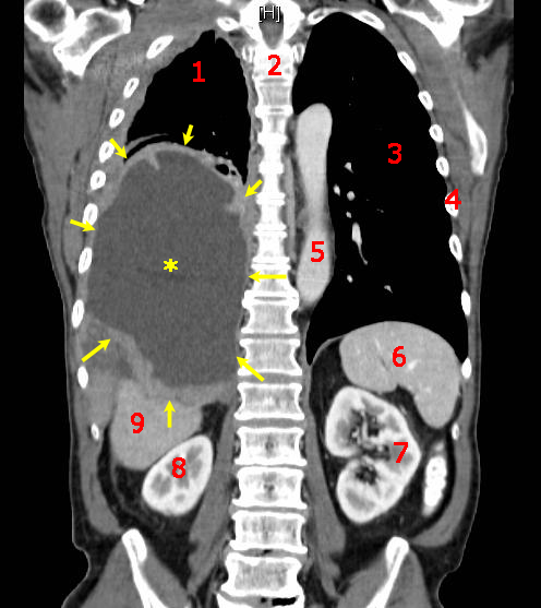
Scan CT (CT scan): procedure whereby a series of detailed pictures of the chest and abdomen is taken from different angles. The images are created by a computer connected to an x-ray machine. Inject a dye into a vein or swallowed to organs or tissues stand out more clearly. This procedure is also called computed tomography, computed tomography or CT scan.
Biopsy: removal of cells or tissues of the pleura or the peritoneum performed by a pathologist to observe them under the microscope and determine if there are signs of cancer.
The procedures used to collect cells or tissues are as follows:
Lung (AAF) fine-needle aspiration biopsy: removal of tissue or fluid using a thin needle. A procedure of images is used to locate abnormal lung tissue or fluid. You can make a small incision in the skin where the needle biopsy to abnormal tissue or fluid, is introduced and a sample is taken. Expand
Thoracoscopy: procedure for which an incision (cut) is made between two ribs and a thoracoscope (a thin instrument tube with a light and a lens for viewing) is inserted into the chest.
Thoracotomy: incision (cut) which is made between two ribs to examine the inside of the chest to determine if there are signs of disease.
Peritoneoscopia: procedure for which an incision (cut) is made in the abdominal wall and a peritoneoscopio (a thin instrument tube, with a light and a lens for viewing) is inserted in the abdomen.
Laparotomy: procedure for which an incision (cut) in the wall of the abdomen is done to check the presence of signs of disease on the inside of the abdomen.
Open biopsy: procedure for which a skin incision (cut) is made to expose and remove tissues in order to examine them and determine if there are signs of disease.
The following tests may be performed on samples of cells and tissues taken:
Cytological examination: examination of cells under a microscope to determine if anything is abnormal. In the case of mesothelioma, removed fluid from the chest or abdomen. A pathologist reviews these liquids for signs of cancer.
Immunohistochemistry: test to which antibodies are used in search of certain antigens in a tissue sample. Antibody is usually linked to a radioactive substance or a dye that makes that tissue illuminates under a microscope. This type of test can be used to tell the difference between different types of cancer.
Electron microscopy: laboratory test in which there are cells of a sample of tissue under the microscope of high power in order to see changes in the cells. An electron microscope shows better than other types of microscopes tiny details.
Certain factors affect prognosis (chance of recovery) and treatment options.
The prognosis (chance of recovery) and treatment options depend on the following:
The stage of the cancer.
The size of the tumor.
If the tumor can be removed completely by surgery.
The amount of fluid in the chest or abdomen.
The age of the patient.
The degree of activity of the patient.
The general State of health of the patient, including the health of the lungs and the heart.
The cell type of mesothelioma and its appearance under a microscope.
The number of white blood cells and the amount of hemoglobin in the blood.
If the patient is male or female.
If the cancer has just been diagnosed or has recurred (come back).
No comments:
Post a Comment